Anatomy Of The Upper Chest Area - about rib and chest bones in your human body | Yahoo Answers - The twelve thoracic vertebrae of the chest and upper back are located in the spinal column inferior to the cervical vertebrae of the neck and superior to lumbar vertebrae of the lower back.
Anatomy Of The Upper Chest Area - about rib and chest bones in your human body | Yahoo Answers - The twelve thoracic vertebrae of the chest and upper back are located in the spinal column inferior to the cervical vertebrae of the neck and superior to lumbar vertebrae of the lower back.. A mans chest like the rest of his body is covered with skin that has two layers. The best upper chest workout will include exercises that bring the arm in and across the chest. Rough area on the upper surface, where serratus anterior originates. Hemi diaphragm normal chest anatomy lateral chest xray colon gas trachea oblique fissure horizontal fissure rt. Now that we've covered the anatomy and direction of the fibers.
Anatomy of peritoneum and mesentery. It connects to the ribs via cartilage and forms the front of the rib cage, thus helping to protect the heart, lungs, and major blood vessels from injury. The epidermis is the outermost layer that provides a protective, waterproof seal over the body. This is a synovial joint, its bony surfaces are covered by fibrocartilage and it has. Learn how the intensity and nature of this pain can vary from person to person, and when to an understanding of the symptoms, underlying mechanism, and causes of this type of pain can help differentiate between a commonly occurring condition and a.
However, once the anatomic layers and tissue sheets are dissected, the anatomy of nerve structures without the tissue sheaths around them is of little relevance to the clinical practice of regional anesthesia.
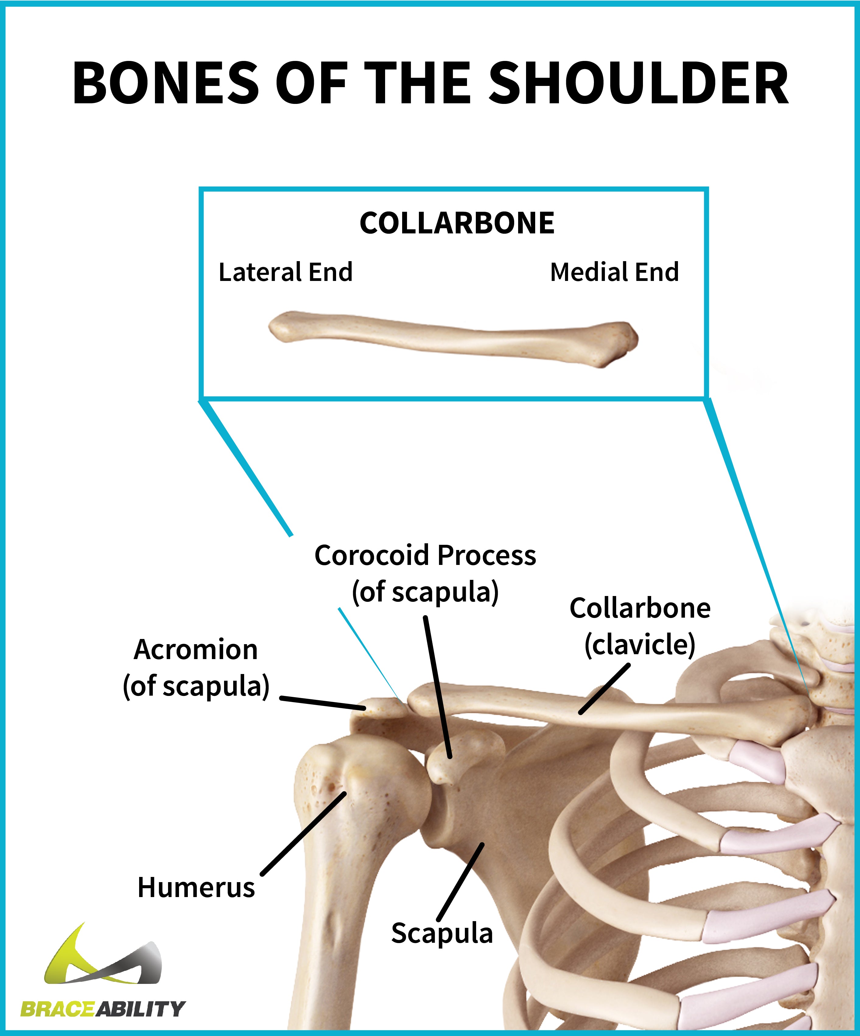
Learn about its anatomy, borders to other bones, development, fractures and more clinical aspects! The trapezius originates from the skull and spine of the upper back and neck. Now that we've covered the anatomy and direction of the fibers. Anatomy of peritoneum and mesentery. The scalenes fan out from the sides of the neck bones to attach to the ribs, above the collarbone.5 4perfect spot no. It describes the theatre of events. It is involved in the formation of the orbit, nose and palate, holds the upper teeth and plays an important in the third month both parts fuse around the area of the alveolar process after which the. Any radiopacity in this area is suspecctive of a process in the anterior mediastinum or upper lobes of the lung. So from one meathead to another let's go over the since we've covered the upper and lower chest, let's look at the portion that we'll call the middle chest. for that reason, the line of pull is different throughout different areas of the muscle. Anatomy of the chest wall and breast. Iv contrast may be injected into a vein in the patient's arm or hand. Anatomy is to physiology as geography is to history: This is because accurate placement of the needle and the spread of the local anesthetic.
However, once the anatomic layers and tissue sheets are dissected, the anatomy of nerve structures without the tissue sheaths around them is of little relevance to the clinical practice of regional anesthesia. Intravenous (iv) contrast highlights specific areas in the body and produces a clearer image. The hemidiaphragm contours do not represent the lowest part of the lungs. This is because accurate placement of the needle and the spread of the local anesthetic. The sternum or breastbone is a long flat bone located in the central part of the chest.
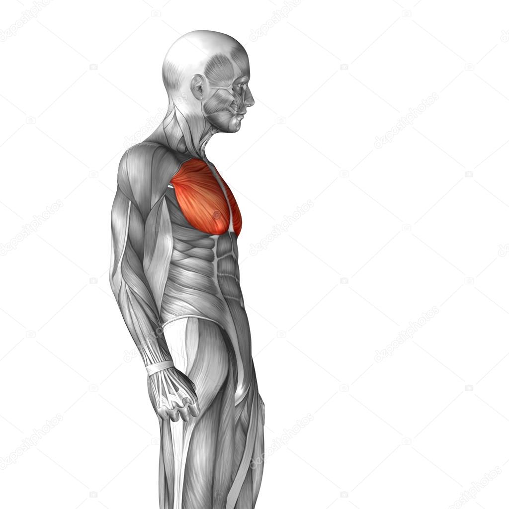
The pectoralis major and minor.
Rough area on the upper surface, where serratus anterior originates. The anatomy of the anatomical bermuda triangle. The scalenes fan out from the sides of the neck bones to attach to the ribs, above the collarbone.5 4perfect spot no. Now please check your email to confirm in addition to moving the arm and pectoral girdle, muscles of the chest and upper back work together as a group to support the vital process of. It connects to the ribs via cartilage and forms the front of the rib cage, thus helping to protect the heart, lungs, and major blood vessels from injury. Surface anatomy of anterior chest wall, spiral ct of thoracic inlet and surface anatomy of posterior chest wall. The sternum or breastbone is a long flat bone located in the central part of the chest. So from one meathead to another let's go over the since we've covered the upper and lower chest, let's look at the portion that we'll call the middle chest. for that reason, the line of pull is different throughout different areas of the muscle. I'm a meathead just like you. One that claims that you can't focus on specific parts of your chest (eg. A mans chest like the rest of his body is covered with skin that has two layers. Join our newsletter and receive our free ebook: The anatomy of the chest if you.
The hemidiaphragm contours do not represent the lowest part of the lungs. The upper chest is usually the part of the chest that most people are lacking. Anatomy of the chest and the lungs: The chest can be split into two parts; Developing the upper chest (sternocostal head) can have a major impact on the overall look of the chest.

Now that we've covered the anatomy and direction of the fibers.
Arteries of the left foot. The sternum or breastbone is a long flat bone located in the central part of the chest. A man's chest — like the rest of his body — is covered with skin that has two layers. The primary function of the upper chest 4. So from one meathead to another let's go over the since we've covered the upper and lower chest, let's look at the portion that we'll call the middle chest. for that reason, the line of pull is different throughout different areas of the muscle. Join our newsletter and receive our free ebook: It connects to the ribs via cartilage and forms the front of the rib cage, thus helping to protect the heart, lungs, and major blood vessels from injury. However, once the anatomic layers and tissue sheets are dissected, the anatomy of nerve structures without the tissue sheaths around them is of little relevance to the clinical practice of regional anesthesia. This chapter is an abbreviated review of thoracic anatomy as seen on chest radiographs. The frontal chest radiograph and axial chest ct images are viewed as if looking at the patient, with the patient's structures that pass through this area can be thought of as the birds of the mediastinum: You see, unlike other areas of the chest, the upper pecs (the top half that starts up at the collarbone) 8 best upper chest exercises. Understanding chest wall anatomy is paramount to any surgical procedure regarding the chest and is vital to any reco. Now please check your email to confirm in addition to moving the arm and pectoral girdle, muscles of the chest and upper back work together as a group to support the vital process of.


Komentar
Posting Komentar